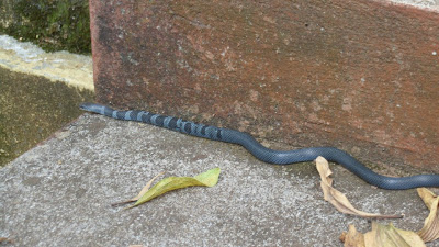
If you need to identify a snake, try the Snake Identification Facebook group.
For professional, respectful, and non-lethal snake removal and consultation services in your town, try Wildlife Removal USA.

Wednesday, December 26, 2012

The Unusual Soft Anatomy of Snakes


This year I had the opportunity to help my friend Lori Neuman-Lee dissect a number of Wandering Gartersnakes (Thamnophis elegans) for her research on the effect of toxic chemicals on reptile physiology. During my Master's, I had the opportunity to help teach a Comparative Anatomy course, during which I learned a great deal about the internal anatomy of vertebrates. However, dissecting an animal for research requires greater accuracy and precision than dissecting for teaching, and we decided to read up on snake anatomy before we got started. Because we found few resources to aid us in our work, we video-taped one of the dissections to help future would-be snake anatomists locate and identify snake organs, several of which can be a little tricky. Check out the video below and learn to dissect a snake! Lori is doing the dissection in the video, and like many things, she makes it look easy. I would recommend some pretty intense practice first if you want to become as accomplished as she is. Salvaged, all-too-common road-killed specimens often make for ideal practice if you don't mind bits of them being smashed, and they sometimes have interesting things in their stomachs.


A few notes: Snakes are long - it's in the blog title. But the implications of being long for the internal anatomy of an animal are not usually considered. For example, in humans and most other animals, paired organs, such as kidneys, lungs, and gonads, are found next to one another, across the body's plane of symmetry. This is not so in snakes; there simply isn't room. Add to their body shape the fact that a great deal of the body cavity must sometimes be filled with eggs or prey items, and there's little room left for the vital organs: heart, lungs, liver, kidneys, spleen and pancreas. That's why snakes have A) evolved elongate organs, B) evolved staggered paired organs, and C) lost some organs or members of paired organs.


Many snake organs are similar in shape to their overall body form. The liver, stomach, gonads, kidneys, and lung are all elongate. Those that come in pairs are either staggered, such as the kidneys and gonads (right always anterior to left), or asymmetrical, such as the lungs. See the tiny left lung near the heart? In a real snake it's almost impossible to find. It is a vestigial organ, meaning it does not function in breathing any more, although in some sea snakes it does have a co-opted function: it helps regulate buoyancy much like the swim bladder of a fish. These adaptations are part of what makes snakes so amazing and unique.

Finally, because 2013 has been designated the Year of the Snake by non-profit conservation group Partners in Amphibian and Reptile Conservation, I hope to help them promote snake research and snake conservation through frequent writing and outreach. As always, thanks for your comments and your readership. Life is Short but Snakes are Long received over 22,000 hits in 2012 and I'm looking forward to an even bigger 2013!


Creative Commons License

Life is Short, but Snakes are Long by Andrew M. Durso is licensed under a Creative Commons Attribution-NonCommercial-NoDerivs 3.0 Unported License.

Monday, December 10, 2012

Magnificent Meizodon


It isn't often that I'm sent a photo of a snake that I can't identify, but last week Alvaro Pemartin found the time both to finish translating all of my old blog posts into Spanish (many thanks to him!) and to obtain photographs of a snake that I had never heard of before. When he sent them to me, I was struck by how beautiful and distinctive the snake was, and it turned out to be quite difficult to identify. I shouldn't have been surprised, because it is from a herpetologically-poorly-known region of the world, western Africa. Alvaro works as a doctor in Guinea, and his nurse, Sandrine Chabassieu, photographed the snake as it crawled across their porch one day.

The mystery snake
Although Kate Jackson's new key to identifying snakes of western and central Africa is a great tool, we couldn't use it to identify this snake because we didn't have close-up pictures of the all-important scale characters that can be indispensable in identifying species of snakes. The backup method, sending the photos to as many people who might know as possible, eventually proved effective when Laurent Chirio, of the Muséum National d'Histoire Naturelle in Paris, France, wrote me that "This is without any doubt a beautiful Meizodon coronatus. I found some specimens with this kind of colour pattern in Guinea."

I wanted to learn what I could about this snake, because it was so striking and previous unknown to me. There isn't a lot out there. Meizodon coronatus, also known as the Western Crowned Snake, is one of five species in Meizodon, a genus of poorly known colubrine snakes found in sub-Saharan Africa. Western Crowned Snakes are found from Senegal to the Congo along the coast of western Africa, while the other four species are found in eastern and central Africa. The Western Crowned Snake was the first species of Meizodon described, by Hermann Schlegel in 1837, who called it Calamaria coronata in his first major published work (Essai sur la Physionomie des Serpens) as assistant curator of the Natural History Museum of Leiden in the Netherlands. The species was later moved to Coronella (subgenus "Mizodon") by Giorgio Jan of the Milan Museum in 1866, then placed in Meizodon in 1955 by Arthur Loveridge.

Plate from Jan 1866

No genes of any species of Meizodon have been sequenced, so it's difficult to say exactly how they are related to other snakes, but if their placement in Colubrinae is correct, then they are probably closely related to other west African species of colubrine, such as the egg-eating Dasypeltis and wolf-toothed Lycodon. These snakes are descended from the same common ancestor as North American kingsnakes and ratsnakes, from which they diverged about 33 million years ago.

Another of Sandrine's photos

Meizodon are primarily predators of lizards, especially skinks, although they are also known to eat geckos, small mammals, and frogs. They are not as specialized for feeding on skinks as their relatives the wolf snakes (Lycodon), on which an upcoming article will focus, but their genus name describes their teeth, which increase in robustness toward the back of the jaw. Meizodon coronatus seem to be a diurnal foragers. A few individuals were observed foraging along the base of a crumbling wall by Godfrey Akani and colleagues at Rumueme, Nigeria. These snakes probed with their heads into holes and crevices where geckos and other lizards were sleeping. Typically, Meizodon are apparently found in refugia such as under rocks, within hollow trees, and underneath loose bark during the day. Don Broadley of the Zimbabwe Natural History Museum  recalls capturing several Meizodon semiornatus that were sheltering inside tree hollows in a flooded forest in western Botswana, along with the psammophine snakes Psammophylax and Psammophis. Broadley noted that several snakes were sometimes captured per tree, including evidence of shed skins and skeletons, although these observations might be atypical given the flooded nature of the forest at the time of Broadley's visit.

The best of Sandrine's photos, in my opinion, showing the gorgeous anterior pattern.

Likely viviparous, Meizodon coronatus inhabits savannahs, forests, plantations, and urban areas. They are mentioned in a study conducted by Godfrey Akani and colleagues on anthropogenic causes of snake mortality in west African suburbs. They found that anthropogenic snake mortality in suburbs of southeastern Nigeria were about 50% intentional, 50% unintentional (e.g., roadkills, snares set to trap more edible wildlife), and that more snakes were killed in the wet season, when they are presumably more active. Even though most of the snake species in this region are harmless and beneficial to humans (in that they exert strong top-down control on populations of pesty rodents, by eating them), most people did not know how to differentiate venomous from non-venomous snakes. It's interesting to know that some ecological problems are common to places as different as North America and Africa. I was glad to hear from Alvaro and Sandrine that, now that they know their beautiful snake is a harmless Meizodon, they have encouraged their camp guards not to kill other Meizodon they might see in the future.

M. coronatus from Cameroon. Not nearly as striking as the one from Guinea.
Edit: 4-Dec-2019: In response to a claim that Meizodon are opisthoglyphous, I revisited some of the literature and found that the original description of Meizodon coronatus by Schlegel in 1837 states (p. 47):
Elle a les dents toutes d'égale longueur ("The teeth are all of equal length")
Günther (1860, p. 429) wrote of M. coronatus that:
The maxillary teeth form one continuous series; anteriorly small, they gradually become longer and stouter posteriorly; none of them are grooved.
Oberkiefer mit 18-19 Zähnen; die ersten sind sehr klein, dicht gedrängt; sie werden nach hinten etwas grösser und weitläuftiger, so dass die letzten doppelt so gross sind als die ersten; ohne Lücken, Keiner derselben ist gefurcht. Unterkiefer mit 20-22 Zähnen, von denen die ersten wenig grössen sind als die letzten, und nach hinten allmählich an Grösse abnehmen. Gaumenzähne: Palatini 12, pterygoidei 14-16, die letzten schwach nach innen gekrümmt (Upper jaw with 18-19 teeth; the first ones are very small, densely crowded; they become slightly larger and wider towards the back, so that the last ones are twice as big as the first ones; without gaps; none of them are grooved. Lower jaw with 20-22 teeth, of which the first are slightly larger than the last, and gradually decreasing in size towards the back. Palatine teeth: 12, pterygoid teeth 14-16, the last teeth slightly curved inwards)
Although the genus name Meizodon comes from the Greek words meizo ("larger" or "greater") and -don (from odonto = "tooth"), this seems to refer to a gradually increasing series rather than an enlarged rear tooth or pair of teeth. I was only able to find a single illustration of the teeth of these snakes (here and above; reprinted from Jan 1866 as part of a detailed comparison of M. regularis and M. coronatus written in 1969). Although there are opisthoglyphous colubrine colubrids (e.g. Dispholidus, Thelotornis) that can kill humans, most species, even those with enlarged rear teeth, do not possess venom dangerous to humans (although colubrid venom and its relationship to tooth anatomy is still on its way to becoming well-understood). Eventually the skull of a Meizodon will be CT scanned & posted here, but until then the single drawing from 1866 and the written descriptions are all we have to go on.

ACKNOWLEDGMENTS

Thanks to Alvaro and Sandrine for the photos, and to Pierson Hill, Peter Uetz, and Laurent Chirio for nailing the ID.


REFERENCES

Akani G, Eyo E, Odegbune E, Eniang E, Luiselli L (2002) Ecological patterns of anthropogenic mortality of suburban snakes in an African tropical region. Isr J Zool 48:1-11 <link>

Barbault R (1976) Population dynamics and reproductive patterns of three African skinks. Copeia 1976:483-490 <link>

Böhme W (2000) Diversity of a snake community in a Guinean rain forest (Reptilia, Serpentes). Bonn Zool Monogr 46:69-78

Broadley DG (1988) Meizodon semiornatus semiornatus: Semiornate Snake. Habitat, Diet, and Distribution. The Journal of the Herpetological Association of Africa 34:44

Günther A (1860) On a West-African genus of snakes (Meizodon). Proc Zool Soc Lond 28:427-430

Jan G (1866) Iconographie Générale des Ophidiens. Livraison. J.B. Bailière et Fils, Paris <link>

Luiselli L, Akani GC, Angelici FM (2001) Diet and foraging behaviour of three ecologically little-known African forest snakes: Meizodon coronatus, Dipsadoboa duchesnei and Hapsidophrys lineatus. Folia Zool 50:151-158

Schlegel H (1837) Essai sur la physionomie des serpens. Partie descriptive. Kips and Van Stockum, La Haye

Segniagbeto GH, Trape JF, David P, Ohler A, Dubois A, Glitho IA (2011) The snake fauna of Togo: systematics, distribution and biogeography, with remarks on selected taxonomic problems. Zoosystema 33:325-360

Roux-Estève, R. 1969. Étude comparée de Meizodon coronatus (Schlegel) et de Meizodon regularis Fischer (Colubridés - Serpentes). Bull. Mus. Natl. Hist. Nat. Paris (ser. 2) 41: 395-409


Creative Commons License

Life is Short, but Snakes are Long by Andrew M. Durso is licensed under a Creative Commons Attribution-NonCommercial-NoDerivs 3.0 Unported License.

Tuesday, November 27, 2012

Snakes of Western and Central Africa


West-Central Africa is, herpetologically, a little known region of the world, although the herp biodiversity there is high. My friend Kate Jackson is one of the few herpetologists to have worked in the region, which you can read all about in her book Mean and Lowly Things. Recently I learned that, in the course of her fieldwork in the Congo, Kate was the first person to photograph a live Bothrolycus ater (Günther’s Black Snake), a rare species of lamprophiid known from only a few specimens collected in Cameroon, Equatorial Guinea, Congo, and Gabon. I was all ready to do a whole post about Bothrolycus when I learned that Darren Naish over at the wonderful blog Tetrapod Zoology had scooped me! This is the first time this has happened because, as Darren has repeatedly pointed out, there are far too few popular snake articles out there.

Kate's Bothrolycus ater picture
But I thought I would take an opportunity to highlight some new resources that are becoming available on west African snakes, in part because a new partner of Life is Short but Snakes are Long, Dr. Alvaro Pemartin, is a Remote Site Doctor working in Guinea. Dr. Pemartin wrote me last week to offer to help translate my articles into Spanish so that they might reach a wider audience. Many many thanks to him for this generous offer! Coincidentally, for people like Dr. Pemartin working to treat snakebite in west-central Africa, Kate Jackson and her students have unveiled a new key that can be used to identify snakes in this region to genus using characters like those I highlighted in my snake sheds post in order to determine the proper kind of antivenom to use. A companion book, Snakes of Western and Central Africa, by Jean-Philippe Chippaux and Kate Jackson, will be available in 2013 from Johns Hopkins University Press (including chapters reviewed by yours truly).

Snakebite is a serious health issue in parts of the developing world, but in North America, it's really a very minor issue. Treatment has advanced to the point where a venomous snakebite, while unpleasant and to be avoided at all costs, is no longer life-threatening unless you are immune compromised. About 5 people a year are killed by venomous snakes in North America, on the order of the same number killed by fireworks. Far more people are killed each year by almost any other cause of death you care to name. Snakes bite people in defense, not in offense. Experiments have shown that venomous snakes 'meter' their venom, often electing not to inject any when biting defensively, and that they often don't bite at all unless severely harassed first. This makes sense, since snakes need their venom to incapacitate their prey and don't want to waste it on predators. The best way to avoid venomous snakebite is to avoid initiating contact venomous snakes. You can be sure that they will avoid you. In case you're afraid of snakes, check out this PSA from The Orianne Society highlighting the many ways snake venom is used to make pharmaceuticals and treat heart attacks, strokes, and cancer; it might make you feel differently.


Today only, donations to the Orianne Society will be matched.

For more on African snakes (man-eating pythons this time), see Emily Taylor's latest post at Ophidiophilia.

ACKNOWLEDGMENTS

Thanks to Kate Jackson for her photos.

REFERENCES

Clarke, DN, Kunkel, W, Chippaux, JP, and Jackson, K. 2012. Online multivariate key to the snake genera of Western and Central Africa. http://people.whitman.edu/~clarkedn/


Creative Commons License

Life is Short, but Snakes are Long by Andrew M. Durso is licensed under a Creative Commons Attribution-NonCommercial-NoDerivs 3.0 Unported License.

Sunday, November 18, 2012

Identifying snake sheds, part III


I noticed that a huge proportion of the hits on this site are for the posts about identifying snake sheds (parts I and II), which I expect is a result of people searching for a key or guide to use to ID a snake shed that they have seen or found. Even though there is some useful information in those other posts, they are written more like detective stories with a particular conclusion in mind, and they certainly aren't comprehensive.

Here, however, I've attempted to put together a more complete how-to guide on how to ID sheds of snakes found in the United States and Canada. One excellent free reference on this subject is an electronic pamphlet by Brian Gray called A Guide to the Reptiles of Erie County, Pennsylvania. Even if you don't live in Erie County, Brian's section on shed snake skins is a very useful guide to many of the common species found in the eastern United States, because it contains many excellent, high-resolution images of the scale characters, and it is organized as a dichotomous key: a series of questions, each with two choices, that inevitably leads to an identification (it's sort of like a choose-your-own-adventure book). Brian's more comprehensive book, The Serpent's Cast, is also an excellent resource, containing images of shed skins that have been painstakingly prepared for viewing the details of the scales necessary for identification to species. Although shed skins that you are trying to identify won't always be that cleanly preserved, often many of the identifying features are still visible.

From Cardwell 2011; viper (top) and colubrid (bottom)
The first thing that many readers will want to know will be whether or not the snake whose shed skin they have found is a venomous species. This distinction corresponds nicely with determining what family the snake is in. In most of North America, there are two families: Viperidae (vipers, which are venomous) and Colubridae (colubrids, which are not1). The easiest way to distinguish these two families by their shed skins is to locate the sub-caudal scales (the scales under the tail). Colubrids have a double row of scales under the tail, whereas vipers have a single row. This is a pretty invariant character2, especially near the anterior part of the tail, and it can help you tell the family of the snake whose shed you've found every time. Coral snakes, which are in the family Elapidae, also have a double row of scales under the tail, but if you think you have found a coral snake shed, post a pic because that's an amazingly lucky find. More about these, and a few other options, later. First, colubrids:

Divided anal scale
Single anal scale
Once you have figured out the family, a second pair of characteristics can help you narrow down which genus of colubrid you might have. These are 1) the texture (smooth or keeled) of the dorsal scales (these are the relatively small scales that cover the snake's entire back and sides) and 2) the condition (single or divided) of the anal scale or anal plate (the scale covering the cloaca). Keeled dorsal scales have a ridge running down the center, whereas smooth dorsal scales have no ridge, like so:

Smooth (left) and keeled (right) dorsal scales
Using these characteristics in tandem should allow you to divide the colubrids in to four groups: single/smooth, divided/smooth, single/keeled, and divided/keeled. These are not taxonomic groups (that is, not all single/smooth snakes are each others' closest relatives), but they are useful for distinguishing genera of colubrids when all you have to go on is the shed skin. All North American vipers have keeled scales and a single anal scale, so these characters are less useful for distinguishing them, but more on these later. Most of the species of North American snake are colubrids (about 80%, or 105 of our 131 species). Here is a quick guide to the colubrids of the US and Canada, by dorsal and anal scale characteristics:


A few genera are split among multiple categories: Gyalopion because G. quadrangulare has a single anal scale whereas G. canum has a divided anal scale, and Opheodrys and Virginia because one species of each has keeled scales and the other has smooth (these are helpfully called Rough and Smooth Green and Earth Snakes, respectively). It's also worth noting that anal scales of Farancia are pretty variable, although your chances of finding a Farancia shed are slim (but see part I).

As you can see, we are using the process of elimination to narrow down the possible candidate species for your shed. A quick look at the range maps in a regional field guide will allow you to cross off about half the genera on the above list, depending on where you live, probably leaving you with 2-6 possibilities. The overall size of the shed can also be of help, although keep in mind that large snakes are born small and that snake sheds stretch somewhat as they are removed. Still, many of the snakes on the above chart reach adult sizes of only 12-24", so they could potentially be eliminated on the basis of size. Width of the ventral scales can help too, because it gives you an idea of body shape, and this does not change as much during the shedding process. However, at this point, the most useful thing to do next is to look at another scale meristic. One that can help you distinguish among the several genera within each group requires counting the dorsal scale rows. Dorsal scales are arranged in rows, the number of which can be counted from left to right, like so:

Three equally good ways to count dorsal scale rows (in C, scale 1 not shown). Modified from K. Jackson (2013)
You'll want to start with the first dorsal in contact with a ventral on one side and proceed over the back and down the other side so that the last scale counted is the dorsal scale in contact with a ventral on the other side of the snake. Although the conventional way (A) is for this to be the same ventral scale as the one your first dorsal scale row was in contact with (that is, count in a ‘V’ shape, as depicted above, so that you are counting all the scales associated developmentally with a single pair of ribs), you should get the same result even if your 'V' is asymmetrical (B), or even if you count in a straight line (C), which can be easier since you don't have to decide where to change direction on the 'V'. Often it doesn't matter, although it's worth noting that in some snakes the number of dorsal scale rows varies along the length of the snake. The best way to guard against this is to count a row in the middle of the body, which is the number meant if only one is given in most keys. More often, you will see numbers of dorsal scale rows given in the format “15-17-15”, indicating the number of dorsal scale rows at three places on the body (in order): the neck, midbody, and a bit (about one head length) before the cloaca.

In North America, you should almost always get odd numbers, and although these numbers can sometimes be fairly variable, combining them with decisions you made above based on the subcaudals, anal scale, dorsal texture, body size, and range should allow you to decide on a genus in almost 100% of cases. Here is a list of the dorsal scale formula ranges for the North American colubrids (remember, it's neck, midbody, and before the cloaca). Where ranges are given in parentheses, species within that genus have differing scale formulas. Where ranges are given without parentheses, there is regional or other variation within one or more of the species in that genus. In a few cases, only the scale row counts at midbody are given.


Knowledge of the number, shape, and relative size of the head scales is usually necessary to distinguish among species within a genus (for example, to tell a Scarlet Kingsnake from a Mole Kingsnake), and unfortunately many sheds are missing their heads or the heads are in poor condition. Other clues can be obtained from pattern, which is often visible in good light, and from counting the total number of subcaudal or ventral scales (impossible if you only have a partial shed). If you have taken your shed to genus and want to send me pictures of the head for help identifying it to species, feel free. I would recommend using your digital camera's macro setting (almost all cameras have one, the symbol is a little flower) to photograph snake sheds. You can also find details of the head scalation of all species of North American snakes in the book Snakes of the United States and Canada by Ernst & Ernst, and much of this information is available online as well. It's often helpful to keep the shed in a Ziploc bag for later reference. I like to write on the bag with a Sharpie the date, location, and tentative ID of the snake.

Non-colubrids

As I mentioned above, all North American vipers have single subcaudals, keeled dorsal scales, and a single anal scale, so these characters are less useful for distinguishing them from one another. However, there are only three genera: Agkistrodon (Copperheads and Cottonmouths), which have no rattles, and two genera of rattlesnakes, Crotalus (which have small scales on the tops of their heads) and Sistrurus (which have large scales on their heads). Telling the different species of Crotalus by their sheds could be tricky, but unless you live in Arizona, there are usually only one or two options in any given location in the US. Size and pattern could also be helpful. Feel free to share pictures (remember to use macro). Copperhead and Cottonmouth sheds can be hard to distinguish, but range, size, and habitat can help, as well as the presence or absence of a loreal scale (the scale on the face between but not in contact with either the eye or the nostril), which Copperheads have and Cottonmouths do not.

Micrurus fulvius
If you live in certain parts of the US, there are a few other snakes that aren't colubrids or viperids whose sheds you might find. One familiar group is the elapids, represented in North America by the Coral Snakes. One species is found in Arizona and New Mexico, and the other in the southeastern coastal plain from Texas to North Carolina. I have never seen a Coral Snake shed, but I would imagine that the highly contrasting, distinctly banded pattern would be easily visible. However, these can also be distinguished by their scale characteristics: Micrurus fulvius has smooth dorsal scales in 15 rows and a divided anal plate, and Micruroides euryxanthus has smooth dorsal scales in a 17-15-15 pattern with a divided anal plate. The other US elapid, the Yellow-bellied Sea Snake (Pelamis platurus, found in the Pacific Ocean off southern California) sheds at sea, so unless you are in very unusual circumstances the sheds will not be found. They have smooth scales with a 39-47, 44-67, 33-46 row formula and a divided anal plate.

Lichanura trivirgata
If you live in southern California or the intermountain west, there are two species of temperate boids, the Rubber (Charina) and Rosy (Lichanura) Boas, whose sheds you could find. Boa sheds are very different from those of other snakes. Boas have small, round dorsal scales that are very numerous - Charina and Lichanura have 32-53 and 33-49 dorsal scale rows, respectively, so you should be able to tell a boa shed by the small size and number of dorsal scales. Rubber Boas have blunt tails and specialized head scales, whereas Rosy Boas have long tails and unspecialized head scales, and their ranges do not overlap. If you live in southern Florida, you might find sheds of Boa Constrictors or Burmese Pythons, which you should be able to tell by their huge size, or any number of other exotic snakes (good luck with those).

Rena humilis
Finally, the southwestern US is home to several species of scolecophidian blindsnakes in the genera Rena and Leptotyphlops. These are tiny and have undifferentiated body scales, meaning that all scale rows around the entire body (including the underside) are the same width. They are iridescent and extremely difficult to count, which has given rise to one of my all-time favorite quotes from a scientific paper: "We castigate the ancient lineage that begat Liotyphlops, for it is obviously the worst designed snake from which to obtain systematic data" (Dixon & Kofron 1983). An additional species, Ramphotyphlops braminus, is introduced in Florida, Louisiana, and Hawaii, as well as in many other locations around the world (it's parthenogenetic and so a really good invader because it only takes one!). Blindsnakes shed their skins in a series of rings rather than in a single piece, and they are so small that any sheds found would be unlikely to belong to any other kind of snake and so fairly easy to identify.

Feel free to comment or email with questions or photographs. Happy herping!


1 I am making a distinction between North American snakes that are dangerously venomous to humans (vipers & coralsnakes) and those that aren't (colubrids). Although some species of colubrid snake possess deadly venom, such as boomslangs and twigsnakes, these are not native to North America. Other colubrids, including some North American species such as Hog-nosed Snakes (Heterodon), are venomous in the sense that their Duvernoy's gland secretions are toxic to their prey, but are harmless or nearly so to humans. For a very thorough discussion of this issue, check out the book "Venomous" Bites from Non-Venomous Snakes.


2 Long-nosed Snakes in the genus Rhinocheilus can have a mixture of divided & undivided subcaudal scales.


ACKNOWLEDGMENTS

Thanks to Brian Gray, Jack Goldfarb, and JD Willson for their excellent photographs.

REFERENCES

Cardwell MD (2011) Recognizing Dangerous Snakes in the United States and Canada: A Novel 3-Step Identification Method. Wilderness & Environmental Medicine 22:304-308. <link>

Dixon JR, Kofron CP (1983) The Central and South American anomalepid snakes of the genus Liotyphlops. Amphibia-Reptilia 4:2-4. <link>

Ernst CH, Ernst EM (2003) Snakes of the United States and Canada. Smithsonian Institution Press, Washington D.C. <link>

Gray BS (2011) A Guide to the Reptiles of Erie County, Pennsylvania. Natural History Museum at the Tom Ridge Environmental Center, Erie, Pennsylvania. <link>

Weinstein SA, Warrell DA, White J, Keyler DE (2011) "Venomous" Bites from Non-Venomous Snakes: A Critical Analysis of Risk and Management of "Colubrid" Snake Bites. Elsevier, Amsterdam. <link>


Creative Commons License

Life is Short, but Snakes are Long by Andrew M. Durso is licensed under a Creative Commons Attribution-NonCommercial-NoDerivs 3.0 Unported License.

Saturday, November 10, 2012

Snakes that polish their scales, and why they do it


Psammophis schokari eating a lizard, Phrynocephalus
mystaceus
, in Kazakhstan
I really like these snakes, and they have about them a pretty interesting mystery. In the tribe Psammophiini (in family Lamprophiidae), there are at least 50 species of snake in 8 genera native to Africa, the Mediterranean, the Middle East, and central Asia. They are united by several unusual synapomorphies, the most unique of which is the presence of a morphological feature called the external narial valve. This structure, located in the loreal region between the eye and the nostril, is the outlet of a special nasal gland that secretes fluid containing long-chain fatty acids. The function of this secretion is enigmatic. Some experiments show that it can serve to retard evaporative water loss, and some evidence suggests that some of these molecules could be pheromones used in marking territories. Territoriality is only slightly less non-existent among snakes than herbivory, but according to some it is apparently present among certain psammophines, few of which have been well-studied in the wild. Aside from a very interesting study suggesting that releasing small mammals from competition with large herbivores can indirectly increase the abundance of their snake predators (including Psammophis mossambicus), we don't know much about their ecology, but careful observations have revealed a little about the lives of these intriguing snakes.

Subadult Montpellier snake, Malpolon monspessulanus
The external narial valve was described in 1956 by renowned Russian herpetologist Ilya Darevsky, the second person ever to earn a PhD in the Soviet Union and the discoverer of parthenogenesis and polyploidy in reptiles. Darevsky described the gland from a specimen of the Montpellier snake (Malpolon monspessulanus), and such glands have now been reported from all eight genera in the Psammophini. In addition to the gland, psammophines also share peculiar hemipene morphology - that is, the male reproductive organs are unusually thin and smooth for an advanced snake, most of which possess thick, spiny hemipenes that enable prolonged copulation. Sexual dimorphism is also quite pronounced in many of these snakes, although not of tail length (typically, the tails of male snakes are longer and thicker than those of females). For example, male M. monspessulanus are stout, uniformly colored, and up to 2.3 m long, whereas females are slender, spotted, and reach only 1.4 meters.

Beginning in 1898, the earliest observations of these snakes mention their peculiar behavior. Psammophines press the outlet of their narial valve to their skin and thoroughly apply a coating of colorless, fast-drying valve secretion all over their body. Watch this Malpolon insignitus to get an idea, because it's hard to describe.


This behavior has been variously called self-rubbing, self-polishing, or  grooming, and seems to be present in all species of psammophine observed. Several keepers in Europe have made extensive efforts to acquire and videotape species of psammophines, and self-rubbing has now been documented in seven of the eight genera. More intriguing, conspecific psammophines housed together occasionally rub one another, presumably anointing the other snake with secretion from their narial valve. What could this mean?


Psammophis leightoni from Namibia
Several hypotheses have been put forth to explain this unique and intriguing behavior. To date, none have been sufficiently tested to unequivocality, nor are they mutually exclusive. Prior to the 1970s, the prevailing thought was that, since psammophines generally inhabit arid regions, the gland might aid in salt excretion, evaporative cooling, or water retention. In 1978, William Dunson and colleagues published their work on the histology of the gland, and concluded that it did not contain the specialized cytological features associated with salt secretion in the salt glands of reptiles such as sea snakes and marine iguanas. Dunson also characterized the chemical composition of the secretion for the first time, and suggested that the long-chain fatty acids he found might help retard water loss through the skin.


Dunson tested five Malpolon to see if their dermal water loss was unusually low, and indeed it was, approximately ten times lower than that of Kingsnakes (Lampropeltis getula), although water loss rate varied depending on where in the shedding cycle the snakes were. Malpolon also lost proportionally more water via the mouth and cloaca (and less via the skin) than did other reptiles. Dunson also kept Malpolon without giving them access to water, and they did not lose weight, indicating that they were capable of obtaining all the water they needed from their prey. In another experiment, he showed that dehydrated Malpolon did not secrete salt from their narial valve. He made the interesting observation that several frog species of the genus Phyllomedusa decrease their dermal water loss by wiping lipid secretions from skin glands over the surface of their skin:


Could psammophids be accomplishing the same thing with their narial valve secretions? Dunson did not test whether snakes that had just applied the secretion lost less water than those that had not. The snakes polish themselves frequently, especially after ecdyisis and feeding, so water loss rate could be tracked over time. 

Other mysterious pits have been described from the head scales of psammophines: parietal pits on the top of the head and infralabial pits on the lower jaw, both of which seem to be sporadically occurring. Series of shed skins from the very same snake sometimes show these features and sometimes do not. Because histology is lacking for these features, it is difficult to say what they might represent.


Dipsina multimaculata
Because of the remote areas inhabited by many of these snakes, most studies to date are insufficiently replicated to permit concrete conclusions about the function of the polishing behavior. Furthermore, determining the sex of living psammophines is quite difficult on account of their small hemipenes, so behavioral studies are often hampered by inadequate knowledge of the sex of the animals involved. Observations of captive psammophines suggest that these snakes have complex social behaviors, not the least of which is their tendency to polish one anothers' scales. Could this behavior represent mate guarding? A nuptial gift from males to females of fatty acids to help them avoid water loss during pregnancy? Do these snakes mark their territories? Only replicated, scientific studies will tell; until then, competing hypotheses will continue to wax on and wax off.


ACKNOWLEDGMENTS

Thanks to Heather Heinz for drawing my attention to this fascinating system, to Jane Bugaeva for translating Darevsky's 1956 article from Russian, and to photographers Bernard Dupont, Altyn Emel, Michael & Patricia Fogden, and Jeremy Holden, and videographer Ton Steehouder.

REFERENCES

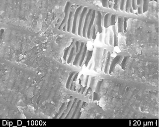
Microdermatoglypic SEM photograph of Dipsina scale.
The lipid layer covering the scale is visible.
From de Pury 2010
Darevsky IS (1956) O stroyenni i funktsionirovani nosovoy zhelezy u yashtsheritsnoy zmei Malpolon monspessulanus Herm. (Reptilia, Serpentes). [On the structure and function of the nasal gland in the lizard snake Malpolon monspessulanus Herm. (Reptilia, Serpentes)] Zoologicheskij Zhurnal-Moskva 35:312-314

Dunson WA, Dunson MK, Keith AD (1978) The nasal gland of the Montpellier snake Malpolon monspessulanus: fine structure, secretion composition, and a possible role in reduction of dermal water loss. Journal of Experimental Zoology 203:461-473

de Grijs P (1898) Beobachtungen an reptilien in der gefangenschaff. Zoologischer Garten 39:233-247

de Haan CC, Aymerich M (2012) Des comportements frotteur et marqueur, pour la chasse et la vie sociale. In: Aymerich M (ed) A la Découverte de la Faune du Maroc Oriental

de Haan CC, A Cluchier (2006). Chemical marking behaviour in the psammophiine snakes Malpolon monspessulanus and Psammophis phillipsi. Proceedings of the 13th Congress of the Societas Europaea Herpetologica, 211-212. <link>

Mimophis mahfalensis killing a chameleon in Madagascar
de Haan CC (2003) Extrabuccal infralabial secretion outlets in DromophisMimophis and Psammophis species (Serpentes, Colubridae, Psammophiini). A probable substitute for ‘self-rubbing’ and cloacal scent gland functions, and a cue for a taxonomic account. Comptes Rendus Biologies 326:275-286. <link>

de Pury S (2010) Analysis of the Rubbing Behaviour of Psammophiids: A Methodological Approach. PhD dissertation, Rheinischen Friedrich-Wilhelms-Universität Bonn, Bonn, Switzerland.

McCauley, D. J., Keesing, F., Young, T. P., Allan, B. F. & Pringle, R. M. 2006: Indirect effects of large herbivores on snakes in an African savanna. Ecology 87, 2657-2663. <link>


Creative Commons License

Life is Short, but Snakes are Long by Andrew M. Durso is licensed under a Creative Commons Attribution-NonCommercial-NoDerivs 3.0 Unported License.

Wednesday, October 3, 2012

Why are there no herbivorous snakes?

This article is part of a series highlighting new research in snake biology presented by herpetologists at the World Congress of Herpetology VII in Vancouver, British Columbia. If you want to learn more about the WCH, check out the June 2012 issue of Herpetological Review, or follow the Twitter hashtag #wch2012, with which I will tag all posts in this series. This article was inspired in part by the research of Beck Wehrle, who did his Master's work on the microbial communities of iguanas, and by Cyndi Carter.


Herbivory (eating plants or their parts) is widespread among vertebrates. There are many herbivorous mammals: think of cows, deer, and other ungulates, as well as lagomorphs (rabbits and their relatives), kangaroos, elephants, sloths, hyraxes, manatees, and even some primates, including many humans. Many birds are herbivorous, including notably the Hoatzin of the Amazon basin, which eats only leaves, but more broadly the many seed- and grain-eating birds such as sparrows, buntings, chickadees, and many other familiar species. Among reptiles, there are herbivorous lizards (including several Filipino monitors, such as Gray's and Panay Monitors, which eat fruit, and Marine Iguanas, which eat only seaweed), turtles (Green Sea Turtles and most tortoises), and even some crocodilians (Simone Brito and colleagues reported Broad-nosed Caimans eating fruit in 2002). Nearly all frogs are herbivorous as tadpoles (and some as adults), and some salamanders, the sirenids, eat algae. There are also many herbivorous fishes: the Pacu of South America, many koi and goldfishes, parrotfishes, and some cichlids and catfishes. Notably absent, then, are the snakes.

A python eats a deer while ignoring plants
There are over 3400 species of snakes, representing >10% of all tetrapods: more than any other group except mammals, birds, and frogs. So why are there no herbivorous species of snake?

At least part of the secret is that nearly all these herbivores (at least, those that eat leaves and parts of plants that have high cellulose content) have something in common: they don't actually digest their own food. You see, much of the energy in plants is stored in their cellulose, a polymer (chain-like molecule) of several hundred to over ten thousand glucose (a common, energy-rich sugar) molecules linked together by β(1,4) bonds. Animals cannot make cellulase enzymes to break the β bonds in cellulose and obtain the monosaccharide glucose, which they can metabolize for energy. Instead, sophisticated endosymbiotic microbes that live in their guts do it for them. The microbes and their herbivorous hosts are digestive symbionts, or indispensable partners. This poses a problem: how do newborn herbivorous organisms get their gut colonies in the first place? The answer: from older members of the same species, including their parents. The transfer is facilitated by processes like live birth, nursing, and coprophagy (eating others' poop), as well as more generally by plenty of social contact thorough pair bonding and parental care, most of which are generally not present in snakes. To be fair, about 15-20% of snakes give live birth, and some have limited parental care and are social at times. But generally speaking, opportunities for microbe transfer between snakes are few and far between. What's more, restrictions of functional morphology probably restrict snakes' ability to evolve herbivory. The kinetic skull with its highly specialized musculature and dentition for swallowing large prey would be extremely unsuitable for the mastication required to pre-digest most plant tissue. There also isn't very much space in a snake's body for the exceptionally long/convoluted gut that seems to be necessary to house the microbial fauna necessary for complete digestion of plant tissue.

Galápagos Marine Iguanas (Amblyrhynchus cristatus)
It's interesting to consider what natural history attributes a herbivorous snake might share with other herbivorous reptiles. Most herbivorous lizards are social, including the Green Iguana and the Marine Iguana. Additionally, herbivory seems to be especially common among lizards on islands, where animal prey  abundance is chronically or periodically low. Predation pressure might also be low on such islands, allowing would-be ectothermic herbivores to meet their energetic demands by eating plants, digestion of which requires prolonged basking to achieve high body temperatures  which could increase predation risk. Many plants reward their herbivores with tasty fruits or nectar in exchange for seed dispersal or pollination - lizards in the MediterraneanNew Zealand, and Mauritius are plant pollinators, and there are lots of examples of mammalian, avian, and invertebrate pollinators. Is it such a stretch to imagine a snake somewhere that enters into this kind of relationship with a flowering plant?

Bagheera
The recent discovery of herbivory in a species of jumping spider, named Bagheera kiplingi by a Jungle Book aficionado,  means that another predominantly carnivorous group contains at least one herbivorous member. These spiders eat Beltian bodies (little packets of fat and protein produced by Acacia trees to reward their ant mutualists/defenders), although they also eat insects. Some other spiders also feed on nectar and might be pollinators. Intriguingly, members of the genus Bagheera are also unusually social for spiders. No word yet on the composition of their digestive microflora, but other plant-tissue-eating arthropods, the termites, rely on similar endosymbioses to vertebrate herbivores.

Tentacled Snake (Erpeton tentaculatum)
Herbivory by a snake has actually kinda-sorta been reported in the peer-reviewed literature once or twice. As far back as 1875, when French naval physician and naturalist Albert Morice wrote the first detailed account of the fauna of Cochinchina, he remarked on how often algae and fragments of aquatic plants were found in the digestive system of the Tentacled Snake, Erpeton tentaculatum. Other authors have remarked on this as well, and according to Morice's account, the local people told him that they knew the snake consumed plants. Hubert Saint Girons, writing in 1972, suggested that in the course of hunting fish in a vegetation-rich freshwater environment, parts of plants might be uprooted and swallowed with the prey. More recent herpetologists have concluded that the presence of plant debris in the stomach contents of E. tentaculatum is, in fact, probably accidental. Furthermore, because there are no  morphological modifications to the dentition or digestive system of E. tentaculatum that would suggest a herbivorous diet, there is no reason to suspect that these plants are consumed on purpose. Nevertheless, the statement that Erpeton is a herbivorous snake has been propagated in several sources, although it is not really accurate.

The more compelling case comes from a study done on island Cottonmouths (Agkistrodon piscivorus) by Harvey Lillywhite and colleagues, published in the journal BioScience in 2008. During a study of pitviper scavenging in the intertidal zone conducted on Florida's Seahorse Key, Lillywhite observed Cottonmouth turds containing relatively large amounts of seaweed (up to 54 grams, or pretty much the entire turd). This seemed like a lot of material to be secondarily or accidentally ingested, and Lillywhite speculated that the Cottonmouths might be eating seaweed because it smelled like fish. Then he and his team went further: to test this idea, they presented Cottonmouths snakes in the laboratory with marine plant materials with and without fish present. The snakes thoroughly investigated the algae lacking fish scent for several minutes, with frequent tongue flicking, pushing, and probing, but they did not attempt to ingest it. When the presentation was repeated using plant materials rubbed with a dead fish or loosely enveloping a piece of fish, the snakes voluntarily swallowed the marine plants that had contacted fish, whether or not the fish was still present. Pics or it didn't happen? Here's the proof:

Figure from Lillywhite et al. 2008

I researched this topic a while ago and intended to make a post about it at some point. What reminded me was actually an experience I had while I was at the WCH: an interesting piece of evidence for a very strange kind of herbivory in a snake came to my attention. My good friend Cyndi Carter, a student of ecology at the University of Georgia, told me I had three chances to guess what she found in a Cottonmouth stomach, and that if I got it she would buy me a large ice cream. My first guess was a dead cat, which they have been known to eat. As a hint, she told me that she found it when she tried to inject the snake with formalin and couldn't get the needle in. After two more wrong guesses on my part (turtle, rock), she showed me this picture:


That's right, a pine cone. Can Cyndi, Joe Mendelson (whose snake it was), or I explain this? We cannot. Maybe it smelled like a fish.


ACKNOWLEDGMENTS


Thanks to Cyndi Carter for the picture of the pine cone, James Van Dyke for articulating what I meant to say about constraints of functional morphology at the end of the third paragraph, Joe Mendelson for clueing me in to the herbivorous frog Xenohyla truncata, and Patrick Prévost for his excellent article and  photograph of Erpeton tentaculatum, one of my favorite snakes.

REFERENCES

Brito, S. P., D. V. Andrade, and A. S. Abe. 2002. Do caimans eat fruit? Herpetological Natural History 9:95-96.

Cowen, R. 1989. Alimentary, My Dear Hoatzin: Ruminations on a Gutsy Bird. Science News 136:269-270.

Dunn, E. R. 1924. Siren, A herbivorous salamander? Science. 59:145.

Farlow, J. O. 1976. Speculations about the diet and foraging behavior of large carnivorous dinosaurs. American Midland Naturalist. 95:186-191.

Fleming, T. H. and K. R. Lips. 1991. Angiosperm endozoochory: were pterosaurs Cretaceous seed dispersers? American Naturalist. 138:1058-1065.

Lillywhite, H. B., C. M. Sheehy III, and F. Zaidan III. 2008. Pitviper scavenging at the intertidal zone: an evolutionary scenario for invasion of the sea. BioScience 58:947-955 <link>

Meehan CJ, Olson EJ, Reudink MW, Kyser TK, Curry RL (2009) Herbivory in a spider through exploitation of an ant–plant mutualism. Current Biology 19:R892-R893. 

Moll, D. and K. P. Jansen. 1995. Evidence for a role in seed dispersal by two tropical herbivorous turtles. Biotropica. 27:121-127. 

Morice, A. 1875. Sur les habitudes d'un remarquable serpent de la Cochinchine: I'Herpeton tentaculatum. Annales des Sciences Naturelles 6:128-129. 

Olesen, J. M. and A. Valido. 2003. Lizards as pollinators and seed dispersers: an island phenomenon. Trends in Ecology & Evolution 18:177-181. 

Pryor, G. S., D. P. German, and K. A. Bjorndal. 2006. Gastrointestinal fermentation in Greater Sirens (Siren lacertina). Journal of Herpetology 40:112-117. 

Sokol, O. M. 1967. Herbivory in lizards. Evolution. 21:192-194. 

Van Damme, R. 1999. Evolution of herbivory in lacertid lizards: effects of insularity and body size. Journal of Herpetology. 33:663-674. 

Walls, G. Y. 1981. Feeding Ecology of the Tuatara Sphenodon punctatus on Stephens Island, Cook Strait. New Zealand journal of ecology 4:89-97.


Creative Commons License

Life is Short, but Snakes are Long by Andrew M. Durso is licensed under a Creative Commons Attribution-NonCommercial-NoDerivs 3.0 Unported License.