If you need to identify a snake, try the Snake Identification Facebook group.
For professional, respectful, and non-lethal snake removal and consultation services in your town, try Wildlife Removal USA.

Wednesday, September 12, 2012

Recent conservation successes with Indigo Snakes

This article is part of a series highlighting new research in snake biology presented by herpetologists at the World Congress of Herpetology VII in Vancouver, British Columbia. If you want to learn more about the WCH, check out the June 2012 issue of Herpetological Review, or follow the Twitter hashtag #wch2012, with which I will tag all posts in this series.


An Eastern Indigo I was very fortunate to see in 2010.
Ask anyone: the Eastern Indigo Snake (Drymarchon couperi) is one of the most commanding and majestic snakes anywhere. Once found throughout Florida and in the coastal plain of southern Georgia, extreme south Alabama, and extreme southeast Mississippi, today the Eastern Indigo survives in numbers only in peninsular Florida and southeast Georgia. Although the species persists in low numbers in the Florida panhandle, it has been extirpated from the rest of its range as a result of declines in and alterations to the longleaf pine (Pinus palustris) ecosystem that it inhabits and on which it critically depends.

One of the largest snakes native to North America, D. couperi is one of five species belonging to a genus that ranges from Georgia to Argentina. Closely related to Racers (Coluber), Patch-nosed Snakes (Salvadora), and Whipsnakes (Masticophis), Indigo Snakes are charismatic and harmless. Carl Kauffeld called them "truly handsome and impressive" in his classic 1957 book Snakes and Snake HuntingMy herpetology professor, Whit Gibbons, told us a story of an exotic dancer who called his lab asking to borrow one to use in her show. This was during the early 1970s, when Eastern Indigos were common in the pet trade, before their federal listing under the Endangered Species Act (one of the first and still one of the only snakes ever listed).

A Gopher Tortoise basks at the entrance of its burrow.
When fire suppression closes the canopy, their basking
and egg-laying microhabitat is lost.
Unfortunately, Indigo Snakes are one of North America's most endangered snake species, primarily as a result of habitat destruction and fragmentation. Research by Natalie Hyslop showed that male Indigo Snakes in southeastern Georgia have home ranges as large as 3,000 acres (nearly five square miles), and one male Indigo Snake moved a distance of about 13 miles (22 km) over two years. As anyone familiar with the southeastern United States knows, it is almost impossible to find five square miles without a road interrupting it, and, as a result, many Indigo Snakes are run over and killed as they cross busy highways and interstates. Conservation of such a highly mobile species is extremely difficult, and by the early 2000s, population strongholds in Georgia were limited to two military bases, Fort Benning and Fort Stewart, where large tracts of uninterrupted sandhill habitat still remain. Furthermore, the degradation of longleaf pine sandhills via fire suppression encourages the growth of hardwood deciduous trees that close the canopy and push out Gopher Tortoises (Gopherus polyphemus), the burrows of which are critical Indigo Snake microhabitats during the winter breeding season. Although habitat degradation is the most insidious factor contributing to Indigo Snake declines, over-collection for the pet trade and malicious killing (both intentional and collateral, as when gasoline fumes are pumped down a tortoise burrow to kill rattlesnakes) are also considerable threats.

In 2008, a non-profit group called The Orianne Society was founded with the purpose of saving the Eastern Indigo Snake from extinction, which seemed inevitable given the rate of land development and habitat degradation in the southeast. TOS has advanced Indigo Snake conservation in a myriad of ways, from acquiring and restoring land to captive breeding. At the Mopani Indigo Snake Preserve in south-central Georgia, TOS biologists are tracking Indigo Snakes using wildlife detector dogs, also used to track other elusive wildlife, from whales to bats to salamanders. Last winter, I was generously invited to witness firsthand the effectiveness of CJ and his handler, biologist Kiley Briggs, at tracking Indigo Snakes at Mopani.

A very happy Orianne Society volunteer holds an Indigo Snake
Indigo Snakes are known to feed primarily on other snakes, lizards, turtles, small mammals, frogs, and birds. Juveniles might feed on fish in the wild, because they spend the early part of their lives in mesic lowland areas and readily consume fishes in captivity. Unusual food items, in comparison to that of other snakes, include small Gopher Tortoises and all venomous snake species native to the Southeastern US (including Copperheads, Cottonmouths, Coral Snakes, and several rattlesnakes). For this last reason, Indigo Snakes generally have a more positive reputation than other snake species among rural residents of the southeast.

Sign alerting motorists to the presence of Indigo Snakes
At the WCH7, Jim Godwin, a zoologist with the Alabama Natural Heritage Program, and Jimmy Stiles, a student with  herpetologist Craig Guyer at Auburn University, brought us good news regarding the Eastern Indigo Snake in Alabama. Due to the collaborative efforts of over a dozen institutions and organizations, including the Alabama Department of Conservation and Natural Resources and TOS, captive-reared Eastern Indigo Snakes have recently been released into Covington County, Alabama's Conecuh National Forest. These snakes were born in captivity from wild females caught in Georgia and head-started at Zoo Atlanta. The plan was to test the effects of a hard (unpenned) or soft (penned) release on snake survival by following snakes with radio telemetry, but the 1 hectare pens built to contain the soft release animals "are just a suggestion to the snakes", according to Godwin. Instead of waiting the intended 90 day soft release period, many of the soft-release snakes released themselves 5-90 days after their initial release, by going under the fence. Because the snakes were implanted with radios, their progress could be followed. Fortunately, the team found that there were no significant differences in survival between snakes that had been hard and soft released, and that hard and soft release snakes had similar sized home ranges. Significantly, the percent overlap between male and female home ranges was higher for soft  release snakes, and this effect increased with time spent in the enclosure. In terms of management implications, releasing snakes in pens does not seem to have a negative effect on snake survival and probably ultimately has beneficial effects on the structure of the established population. Earlier attempts to reestablish Eastern Indigo Snakes in Alabama were unsuccessful, possibly both as a result of the hard release techniques used and the release of too few snakes in too many locations. In the two years since the initial release in 2010, most of the Conecuh Indigos have survived, although several have been killed by predators and several more run over by cars. Improvements in the fire management regime in the Conecuh and continued research on the reintroduced snake population should mean a bright future for the Eastern Indigo Snake in Alabama.

Clearly I could write about Indigo Snakes all day, but if you want to learn more, check out The Orianne Society's website or read some of the papers linked in the References section below.

ACKNOWLEDGMENTS

Thanks to Mark Wallace for his photo of the happy volunteer.

REFERENCES

Bauder JM, Macey JN, Wallace MP, Snow F, Safer AB, Stevenson DJ (2012) Drymarchon couperi (Eastern Indigo Snake). Juvenile observations. Herpetological Review 43:343

Breininger D, Bolt ML, ML, Drese J, Stolen E (2011) Factors influencing home-range sizes of Eastern Indigo Snakes in central Florida. Journal of Herpetology 45:484-490 <link>

Breininger DR, Mazerolle MJ, Bolt MR, Legare ML, Drese JH, Hines JE (2012) Habitat fragmentation effects on annual survival of the federally protected eastern indigo snake. Animal Conservation 15:361-368 <link>

Godwin J, Wines M, Stiles J, Stiles S, Guyer C, Rush EM (2011) Reintroduction of the Eastern Indigo Snake (Drymarchon couperi) into Conecuh National Forest. State Wildlife Action Grant Report. <link>

Hyslop NL, Cooper RJ, Meyers JM (2009) Seasonal shifts in shelter and microhabitat use of Drymarchon couperi (Eastern Indigo Snake) in Georgia. Copeia 2009:458-464 <link>

Stevenson DJ et al. (2010) Prey records for the Eastern Indigo Snake (Drymarchon couperi). Southeastern Naturalist 9:1-18 <link>

Stevenson DJ, Ravenscroft KR, Zappalorti RT, Ravenscroft MD, Weigley SW, Jenkins CL (2010) Using a wildlife detector dog for locating Eastern Indigo Snakes (Drymarchon couperi). Herpetological Review 41:437-442

Stevenson DJ et al. (2009) An Eastern Indigo Snake (Drymarchon couperi) mark-recapture study in southeastern Georgia. Herpetological Conservation and Biology 4:30-42 <link>


Creative Commons License

Life is Short, but Snakes are Long by Andrew M. Durso is licensed under a Creative Commons Attribution-NonCommercial-NoDerivs 3.0 Unported License.

Tuesday, September 4, 2012

Snakes that can see without eyes

This article is part of a series highlighting new research in snake biology presented by herpetologists at the World Congress of Herpetology VII in Vancouver, British Columbia. If you want to learn more about the WCH, check out the June 2012 issue of Herpetological Review, or follow the Twitter hashtag #wch2012, with which I will tag all posts in this series. 


Close-up of pit organ of Tropidolaemus subannulatus
Pit vipers have an amazing and little-known ability to see infrared light. They do this by means of their eponymous pits, which are essentially a second pair of eyes located in the loreal region of their face, between the normal (visible light or "lateral") eye and the nostril. Some snakes, such as Emerald Tree Boas (Corallus caninus), have up to forty pits, meaning that in effect they have forty-two 'eyes': two lateral eyes and forty infrared eyes. Pit vipers have just two, but these organs are among the most exquisitely sensitive sensory organs in the animal kingdom. Other animals can also see wavelengths outside of the spectrum of light visible to humans. For example, bees and many birds can see ultraviolet wavelengths, and the complex eyes of mantis shrimp possess at least 16 different photoreceptor types, allowing them to see visible and ultraviolet light with fine sensitivity, as well as polarized light, thought to allows them to see their transparent prey.

Figure from Goris 2011
a) Boa constrictor b) Corallus caninus
c) Python molurus d) Gloydius blomhoffii
Because pit organs are found in snakes as distantly related as boas, pythons, and rattlesnakes, they must have evolved at least three times over the last 125 million years (boas and pythons, believe it or not, are fairly distant relatives). As you can see, the morphology of the pit organ is very different in these three snake lineages. In pit vipers, it is most sophisticated. The pit viper pit organ is made up of three parts: an inner and an outer chamber, separated by a thin membrane. This membrane functions as an "infrared retina", detecting infrared radiation that enters the inner chamber. The inner chamber cannot be seen from outside of the snake's body, but it communicates with the exterior air via a pore located between the eye and the pit. Because the exterior opening of the outer chamber is smaller than the membrane, infrared light sources cast a shadow on the membrane, which are detected as an image by the nervous system. It works a lot like a pinhole camera. The information is processed by the nervous system separately from that gained using the lateral eyes, but all four (in the case of pit vipers) images are integrated in the brain to produce one single coherent image of the environment. It isn't so different from what your brain does when it integrates two slightly different images of the world, each collected by one of your eyes, to produce an integrated image with depth. The neurology of this process in infrared snakes is relatively well understood, although it is hard to imagine processing visual information from more than two sources.

Rather than thinking of the pits as a "sixth sense", what they actually do is to improve the vision of the snake by making use of parts of the electromagnetic spectrum for which there are no color pigments. To envision this, imaging seeing heat (which is the most common source of infrared radiation) as an additional color. In fact, pit vipers can see differences in temperature in both directions - so an object that is colder than its surroundings also become more visible to the pit organ. It's like the image of a person holding a caterpillar to the left, except with real colors added also. Check out this site for more infrared images.

Innervaton of the crotaline pit organ.
Figure from Goris 2011
During the World Congress of Herpetology's venomous snake evolution session, Bruce Young of the University of Massachusetts at Lowell presented amazing new results revealing directional asymmetry of the thermal image. It was known that, depending on the habitat of the species, there was some difference in the configuration of the pit, but Young's recent work showed that the area of maximum focus (analogous to the fovea of the visible-spectrum eye) is above and behind the head in terrestrial species, and below and behind the head in arboreal species. Because the many uses of the pit organ include enabling snakes to better see predators and prey in great detail in the dark, including those that are partially concealed to the lateral eyes, it could be inferred that these differences in pit organ morphology are determined partially by ecology. Much more work needs to be done on this fascinating system, especially cataloging the diversity of the pit organs of boas (53 species, not all of which have such organs), pythons (41 species), and other pit vipers (216 species).

Cottonmouth (Agkistrodon piscivorus)
When I wrote the title for this post, I realized that it could also apply to two other groups of snakes that get along just fine without eyes. One is the blindsnakes, or scolecophidians, a primitive radiation of snakes about which many fascinating posts are forthcoming. Also worth mention is the population of Tiger Snakes (Notechis scutatus) on Carnac Island in Western Australia. Seabirds, especially Silver Gulls, peck out the eyes of these snakes while defending their nests from predation by the snakes. In a 1999 study published in the journal Behavioral Ecology and Sociobiology, Xavier Bonnet and colleagues found that tiger snakes that had lost their eyes suffered no loss of body condition, growth rate, mating opportunities, or survival. This is especially remarkable because it means these snakes are getting by using tactile and chemosensory information only, since elapids have no pit organ and cannot see infrared light. The late biologist and author extraordinaire Charles Wharton also documented eyeless Cottonmouths on Sea Horse Key in Florida in 1969, which, being vipers, could continue to rely on their pit organs, the function of which was poorly understood at the time.

Tyson's diagram of the head of a rattlesnake;
the pit, which he called the foramen, is at B
Older theories for the purpose of the pits included that they were ears, extra nostrils, organs of smell, secretory organs to wash the cornea, tactile sensors, part of a lateral line system such as that in fishes, or sensory organs of a completely unknown "sixth sense". It wasn't until 1935 that Margarete Ros first associated the pit organs of an African Rock Python with infrared radiation by observing differences in its attentiveness to warm objects before and after she occluded its pits with petrolatum jelly. This was more than 250 years after Edward Tyson first mentioned snake pits at a scientific meeting of the Royal Society of London in 1683, during which he dissected a rattlesnake from Virginia that he called Vipera caudisona (almost certainly a Timber Rattlesnake, Crotalus horridus).

ACKNOWLEDGMENTS

Thanks to Kurt (orionmystery) for his photo of Tropidolaemus subannulatus, and to Pierson Hill for his photo of Agkistrodon piscivorus.

REFERENCES

Bakken GS, Krochmal AR (2007) The imaging properties and sensitivity of the facial pits of pitvipers as determined by optical and heat-transfer analysis. Journal of Experimental Biology 210:2801-2810 <link>

Bonnet X, Bradshaw D, Shine R, Pearson D (1999) Why do snakes have eyes? The (non-) effect of blindness in island tiger snakes (Notechis scutatus). Behavioral Ecology and Sociobiology 46:267-272 <link>

Goris RC (2011) Infrared organs of snakes: an integral part of vision. Journal of Herpetology 45:2-14. <link>

Kohl T, Colayori SE, Westhoff G, Bakken GS, Young BA (2012) Directional sensitivity in the thermal response of the facial pit in western diamondback rattlesnakes (Crotalus atrox). The Journal of Experimental Biology 215:2630-2636 <link>

Safer AB, Grace MS (2004) Infrared imaging in vipers: differential responses of crotaline and viperine snakes to paired thermal targets. Behavioural Brain Research 154:55-61 <link>

Tyson E (1683) Vipera Caudi-Sona Americana, Or the Anatomy of a Rattle-Snake, Dissected at the Repository of the Royal Society in January 1682/3 by Edw. Tyson MD Coll. Med. Lond. Cand. & RS Soc. Philosophical Transactions (1683-1775) 13:25-46 <link>

Van Dyke JU, Grace MS (2010) The role of thermal contrast in infrared-based defensive targeting by the copperhead, Agkistrodon contortrix. Animal Behaviour 79:993-999 <link>

Wharton CH (1969) The cottonmouth moccasin on Sea Horse Key, Florida. Bulletin of the Florida State Museum of Biological Sciences 14:227-272 <link>


Creative Commons License

Life is Short, but Snakes are Long by Andrew M. Durso is licensed under a Creative Commons Attribution-NonCommercial-NoDerivs 3.0 Unported License.

Monday, August 27, 2012

Squirrel v. rattlesnake

This article is part of a series highlighting new research in snake biology presented by herpetologists at the World Congress of Herpetology VII in Vancouver, British Columbia. If you want to learn more about the WCH, check out the June 2012 issue of Herpetological Review, or follow the Twitter hashtag #wch2012, with which I will tag all posts in this series.


It might seem like a lopsided contest, but in the majority of interactions between Northern Pacific Rattlesnakes (Crotalus oreganus) and California ground squirrels (Spermophilus beecheyi), the squirrels walk away with their lives. This surprising result come from Rulon Clark, who in his talk during the venomous snake evolution session of the WCH7 last week filled us in on the latest work from his behavioral ecology lab at San Diego State University. Building on the work done by mammalogists Richard Coss and Don Owings at UC Davis, the Clark lab studies what ground squirrels are trying to say to their rattlesnake predators. You see, when a ground squirrel encounters a rattlesnake, it performs a behavior known as 'tail-flagging'. You can see an example of this behavior in the first half of this video:


and the potential consequence of not exhibiting it in the second half! It's been apparent for almost 35 years now that tail-flagging adult squirrels are safer from rattlesnakes than squirrels that don't perform this behavior, but why?

Dr. Clark enumerated several hypotheses that his lab has tested and falsified:
  • tail-flagging does not appear to be a form of quality advertisement, like stotting in ungulates, because its use is not correlated with the health or vigor of the squirrel
  • tail-flagging does not appear to result in predator confusion or misdirection, because the rattlesnakes that strike at tail-flagging squirrels are equally accurate in their strike direction as those that strike at squirrels that aren't tail-flagging
  • tail-flagging does not appear to be a form of harassment, like mobbing in birds & other animals, because the squirrels never attack rattlesnakes if the snakes are free-ranging (although they will if the snakes are caged, as they were in early experiments) and eventually leave the snakes alone after tail-flagging at them for a while.
Additionally, the tail-flag display is frequently given in the absence of a rattlesnake, as if to probe for potential predators nearby. So how is tail-flagging helpful? By videotaping countless hours of snake-squirrel interactions using stationary cameras - fortunately, rattlesnakes are fairly stationary themselves - Clark's group thinks they have the answer.

Crotalus oreganus from Utah
First, the squirrels are probably advertising their perception of the snakes, both to the snakes themselves and to each other. This is likely because tail-flagging by one squirrel increases the vigilance of other squirrels in the area. Furthermore, rattlesnakes that have been tail-flagged are actually more likely to abandon their ambush sites. Both these things only happen, however, when the tail-flagging squirrel is an adult. Similarly, we respond more seriously to cries of a fire by an adult than by a child. Juvenile squirrels also tail-flag, but presumably they are just practicing, so adults apparently do not take them seriously.

Second, the adult squirrels are probably also advertising their vigilance to the snakes. This is likely for two reasons: 1) the snakes are less likely to strike an adult tail-flagging squirrel than a non-tail-flagging one, and 2) if they do, squirrels that tail-flagged are more likely to successfully dodge the rattlesnake's strike. That's right - these ground squirrels can actually evade the snake's strikes. Don't believe it?


I hardly can either, but wow, that squirrel pulled a 180 and totally avoided what should have been a lethal strike. Although the squirrel in that video wasn't tail-flagging, Clark's group has shown that within about one foot of a rattlesnake, tail-flagging squirrels are more likely to dodge strikes successfully. As a result, rattlesnakes are less likely to strike at a tail-flagging squirrel - not because the energy cost is too high, but because a strike will surely cause the squirrel to run off, while waiting might result in the squirrel making a mistake by getting too close. After all, once a snake has been tail-flagged, it might as well move ambush sites, because the local squirrels are now aware of its presence.

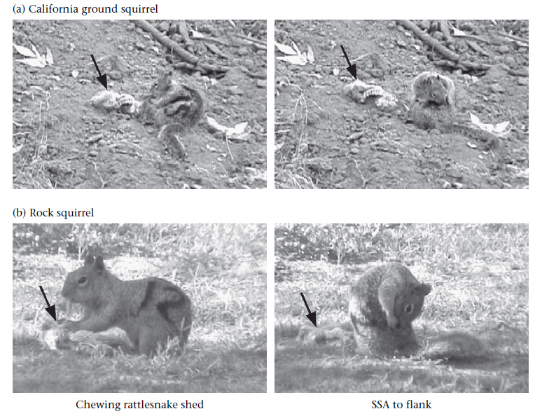
In addition to employing highly effective perception and vigilance advertisement behaviors, those darn squirrels have also evolved to anoint their fur with rattlesnake scent! They get this odor from chewing up shed rattlesnake skins. Barbara Clucas showed that the snake scent application did not deter other squirrels or help reduce ectoparasites, bolstering the case that it is a form of olfactory camouflage that serves to reduce squirrel detectability to snake predators or to repel other rattlesnakes motivated to avoid hunting in the same area as a conspecific.

Figure from Clucas et al. 2008

By now, I imagine the snake biologists in the audience are itching to see a snake actually get one for once. Here you go:


If you want to see more videos and stay current on the Clark lab's research, subscribe to their Youtube channel or to Strike, Rattle, & Roll, a rattlesnake behavior blog published by Clark lab PhD student Bree Putman.

ACKNOWLEDGMENTS

Thanks to Rulon Clark for his helpful review of this article.

REFERENCES

Barbour, M. A. and R. W. Clark. 2012. Ground squirrel tail-flag displays alter both predatory strike and ambush site selection behaviours of rattlesnakes. Proceedings of the Royal Society B: Biological Sciences doi:10.1098/rspb.2012.1112. <link>

Clark, R. W., S. Tangco, and M. A. Barbour. 2012. Field video recordings reveal factors influencing predatory strike success of free-ranging rattlesnakes (Crotalus spp.). Animal Behaviour 84:183-190. <link>

Clucas, B., D. H. Owings, and M. P. Rowe. 2008. Donning your enemy's cloak: ground squirrels exploit rattlesnake scent to reduce predation risk. Proceedings of the Royal Society B: Biological Sciences 275:847-852. <link>

Coss, R. G. and D. H. Owings. 1978. Snake-directed behavior by snake naive and experienced California Ground Squirrels in a simulated burrow. Zeitschrift für Tierpsychologie 48:421-435. <link>

Owings, D. H. and R. G. Coss. 1977. Snake mobbing by California ground squirrels: adaptive variation and ontogeny. Behaviour 62:50-69. <link>

Rundus AS, Owings DH, Joshi SS, Chinn E, Giannini N (2007) Ground squirrels use an infrared signal to deter rattlesnake predation. Proceedings of the National Academy of Sciences 104:14372-14376 <link>


Creative Commons License

Life is Short, but Snakes are Long by Andrew M. Durso is licensed under a Creative Commons Attribution-NonCommercial-NoDerivs 3.0 Unported License.

Tuesday, August 21, 2012

Goo-eating snakes and the eggs that evade them


I have just returned from attending the Seventh World Congress of Herpetology (WCH7) in Vancouver, Canada. This meeting is held once every four years, always in the same year as the Summer Olympics, from which it differs in several important ways. Although many celebrities attend each, the WCH primarily consists of scientific, rather than physical, displays of prowess. Until a gold medal is given in lizard noosing, herpetologists will continue to present their research at the WCH, as I had the opportunity to do this year. Because of the large number of excellent talks highlighting new research in snake biology, I have decided that the next several articles on LISBSOL will constitute a series inspired by the work of the many herpetologists whom I saw presenting at WCH7. If you want to learn more about the WCH, check out the June 2012 issue of Herpetological Review, or follow the Twitter hashtag #wch2012, with which I will tag all posts in this series (disappointing though it is that herpetologists should be forced to 'tweet' their research rather than 'hiss' or 'croak' it [I couldn't figure out how to spell the sound that alligators make]).

One tradition at WCH meetings is to open each day with a plenary talk, which is an hour-long presentation by a distinguished herpetologist. Of the several plenaries at WCH7, the one that impressed me the most was given on the first day by Karen Warkentin, a herpetologist at Boston University who studies environmentally-cued hatching of amphibian eggs. One of the foundations of her research is that the timing of hatching, a critical life-stage transition in the life of an amphibian (or reptile), should be flexible in order to maximize the likelihood of survival of the young animals. That is, if the egg is safe from predators and pathogens, hatching should be delayed as long as possible (typically until the embryo is as large as it can get without leaving the egg). However, if the egg is in danger, hatching should speed up, as long as the embryo is capable of living outside of the egg. This phenomenon is observed in a variety of reptiles and amphibians, including  the Agalychnis (red-eyed) treefrogs that Dr. Warkentin studies. These frogs lay their eggs on leaves overhanging pools in the Neotropical rain forests, so that when they hatch the tadpoles can drop into the water.

Agalychnis callidryas in amplexus
The primary predators of Agalychnis eggs are wasps and snakes. In the wild, snakes consume as much as 50% of all Agalychnis eggs laid, so it makes sense that there would be strong selection for eggs that could escape snake predation. If a snake or wasp attacks a clutch of eggs, the vibrations trigger the eggs to hatch almost immediately. If that sounds impossible, check out this video of a Parrotsnake (Leptophis) attacking a clutch of eggs:


Look at those little guys hatch! You can see other videos at Dr. Warkentin's website, where you can compare the feeding behavior of Leptophis with that of the Cat-eyed Snake (Leptodeira). Embryos in the last third of their development escape from snake attacks with about an 80% success rate by hatching up to 30% early, which is really remarkable. Furthermore, they can distinguish snake attacks from other sources of vibration, so that they don't hatch every time it rains. To do this, they respond to several non-redundant vibrational cues, including frequency, duration, and their interaction. These cues propagate throughout the jelly matrix of the eggs, so that eggs that have not yet been touched by the snake can escape. In two species of Agalychnis that have reduced jelly, escape success is much lower, because the signals do not propagate as well.

Vibration profile of a snake attack

According to Dr. Warkentin, the snakes do not appear to prefer younger eggs (which would be incapable of hatching early) or to forage preferentially in the rain (when their vibrations might be masked by raindrops). Along with Leptophis and Leptodeira, two other snake genera, Sibon and Dipsas, possess morphological and behavioral adaptations for feeding on frog eggs and other prey items that are essentially 'goo'. Not unlike the southeast Asian pareatids I've covered before, these Neotropical snakes have numerous, long, slender teeth on the dentary (lower jaw), and they have many skeletal and muscular modifications that allow for jaw flexibility beyond even that normally seen in snakes. Extinction of many frogs due to chytrid fungus in Central America has caused dietary shifts and changes in abundance of these snakes.

Sibon argus eating frog eggs

Environmentally-cued hatching in response to vibrations also occurs in the eggs of other treefrogs, centrolenid glass frogs, and African reed frogs. It can also occur in response to other environmental dangers, such as flooding (in salamander and some turtle eggs) and disease (in frog eggs and also in painted turtle hatchlings, which often overwinter in the nest but are more likely to emerge early when infected with sarcophagid fly larvae). This last example comes from the thesis work of Julia Riley at Laurentian University, who presented preliminary results at the WCH. She also found that turtles hatching in nests that were on steeper slopes were more likely to emerge early, possibly to avoid collapse of the nest over the winter. Whether research will one day show that snake eggs also possess environmentally-cued hatching plasticity is an open question, but I suggest that a good system to start looking would be the Nicrophorus beetle hosts. Maybe we'll be hearing about that at WCH8 in Hangzhou, China!

ACKNOWLEDGMENTS

Thanks to Otto Monge, Brad Wilson, and the Warkentin lab website for providing photos and videos.

REFERENCES

Caldwell MS, McDaniel JG, Warkentin KM, 2009. Frequency information in the vibration-cued escape hatching of red-eyed treefrogs. J Exp Biol 212:566-575. <link>

Caldwell, M. S., J. G. McDaniel, and K. M. Warkentin. 2010. Is it safe? Red-eyed treefrog embryos assessing predation risk use two features of rain vibrations to avoid false alarms. Animal Behaviour 79:255-260 <link>

Gomez-Mestre I, Warkentin KM, 2007. To hatch and hatch not: similar selective trade-offs but different responses to egg predators in two closely related, syntopic treefrogs. Oecologia 153:197-206. <link>

Gomez-Mestre I, Wiens JJ, Warkentin KM, 2008. Evolution of adaptive plasticity: risk-sensitive hatching in neotropical leaf-breeding treefrogs. Ecol Monogr 78:205-224. <link>

Lips KR, Brem F, Brenes R, Reeve JD, Alford RA, Voyles J, Carey C, Livo L, Pessier AP, Collins JP, 2006. Emerging infectious disease and the loss of biodiversity in a Neotropical amphibian community. Proc Natl Acad Sci USA 103:3165-3170. <link>

Ray JM, Montgomery CE, Mahon HK, Savitzky AH, Lips KR, 2012. Goo-eaters: Diets of the Neotropical snakes Dipsas and Sibon in central Panama. Copeia 2:197-202. <link>

Savitzky AH, 1983. Coadapted character complexes among snakes: fossoriality, piscivory, and durophagy. American Zoologist 23:397-409. <link>

Warkentin, KM, 2005. How do embryos assess risk? Vibrational cues in predator-induced hatching of red-eyed treefrogs. Animal Behaviour 70:59-71. <link>

Warkentin KM, Caldwell MS, McDaniel JG, 2006. Temporal pattern cues in vibrational risk assessment by embryos of the red-eyed treefrog, Agalychnis callidryas. J Exp Biol 209:1376-1384. <link>

Warkentin KM, Caldwell MS, Siok TD, D'Amato AT, McDaniel JG, 2007. Flexible information sampling in vibrational assessment of predation risk by red-eyed treefrog embryos. J Exp Biol 210:614-619. <link>

Warkentin KM, Currie CR, Rehner SA, 2001. Egg-killing fungus induces early hatching of red-eyed treefrog eggs. Ecology 82:2860-2869. <link>


Creative Commons License

Life is Short, but Snakes are Long by Andrew M. Durso is licensed under a Creative Commons Attribution-NonCommercial-NoDerivs 3.0 Unported License.

Scientific American Guest Blog


Check out the piece on toxin-sequestering snakes I was invited to write for the Scientific American Guest Blog!!

http://blogs.scientificamerican.com/guest-blog/2012/08/21/poisonous-snakes-cant-resist-toxic-toad-tucker-or-can-they/

Heterodon platirhinos eating Acris crepitans. Photo by Nick Kiriazis


Creative Commons License

Life is Short, but Snakes are Long by Andrew M. Durso is licensed under a Creative Commons Attribution-NonCommercial-NoDerivs 3.0 Unported License.

Saturday, August 4, 2012

Stiletto snakes


Atractaspis duerdeni
I've always thought that the atractaspids were a highly interesting group of snakes, deserving of an article or two. During the early stages of my cursory research, however, I found that palaeozoologist Darren Naish, author of the excellent blog Tetrapod Zoology, has already written an article containing what one commenter called "the most comprehensive information on Atractaspids anywhere on the web." Since I didn't think I could top that, I decided to focus on what we've learned about atractaspids since Darren's article came out in 2008. If you want to learn more about the many fascinating adaptations atractaspids have evolved for burrowing and closed-mouth fang-stabbing, including why they're known as والد من سواد ('father of blackness'), among other such macabre names, in Arabic-speaking countries in their native range, you'll want to read that article in addition to this one.

The correct placement of the atractaspids within the snake tree of life has been elusive since their initial description in 1843, when they were placed in the Elapidae alongside cobras and coralsnakes. In later classifications, they have been placed in the Viperidae, the Colubridae, the Lamprophiidae, or in their own family, to which various names have been applied, including Atractaspidae (atractaspids), Atractaspididae (atractaspidids, because why not add in an extra 'id'?), and Atractaspidinae (atractaspidines; this last name referring to a subfamily rather than a family). Once considered to include a wider diversity of snakes, the Atractaspidinae is now comprised of just two genera, the proteroglyphous Homoroselaps (2 species, known as Harlequin Snakes) and the eponymous, solenoglyphous Atractaspis (21 species). Aglyphous and opisthoglyphous snakes formerly included in this group are now assigned to a closely related subfamily, the Aparallactinae, which includes 50 species in nine genera, several of which are deserving of their own articles. This taxonomy is based on part of a larger analysis of advanced snakes undertaken by Alex Pyron and colleagues and published in 2010, and hinted at in earlier analyses such as this one by Kraus & Brown.


Part of the tree presented in Pyron et al. 2010, showing the relationships of atractaspids to other African snakes now placed in the Lamprophiidae. A surprising finding of this paper was that lamprophiids share a common ancestor with the front-fanged elapids, including cobras, sea snakes, and coral snakes, about 44 million years ago.
Morphological work on atractaspids has continued to be carried out by Dave Cundall and his students and colleagues at Lehigh University. I had the opportunity to hang out with Dave a bit recently, and he shared some of his recent findings with me. For instance, he said, the long-held idea that Atractaspis fed predominantly on litters of baby mammals might be only party true. The stomach of some atractaspids, he told me, is almost as long as the entire body, an adaptation that could be construed as functioning to accommodate multiple prey items (pups in a litter) but also large, elongate ones (such as amphisbaenians or caecilians), which also frequently occupy underground spaces where hunting by fang-stabbing is effective. Dave also mentioned that digestion in these snakes takes place, as one might expect, only in the stomach, not in the esophagus, although ingested prey may extend forward into the esophagus if they are too large to fit in the stomach. Differences in the tissue lining these two parts of the digestive system account for a pH change of up to 4 units between the esophagus and the stomach, one of the few clues that these two organs in snakes are derived from separate structures in other vertebrates (since their morphological separation in many snakes is subtle at best). Other discoveries made by Dave and his student Alex Deufel, including how atractaspids, perhaps uniquely among advanced snakes, have traded-off prey transport for maximum fang-stabbing ability, have been described in excellent detail by Darren at TetZoo.

No one is quite sure why, but some Atractaspis also possess extremely elongate venom glands, such as those seen here in a dissected A. fallax.

Other recent work on atractaspids includes advances in understanding their unusual venom chemistry and in treating its effects, including the discovery and production of the first atractaspid antivenom in 2007. In a test of this antivenom conducted at the National Antivenom and Vaccine Production Center in Riyadh, Saudi Arabia, rabbits injected with a lethal dose of Atractaspis venom were saved from death by a pre-injection treatment of any one of three drugs: nitroglycerin, atractaspid antivenom, or bosentan, a drug for the treatment of pulmonary hypertension. However, when the drugs were administered after the venom, as would be the case in an actual snakebite, all rabbits treated with nitroglycerin and half the rabbits treated with atractaspid antivenom died. Only the hypertension drug bosentan protected rabbits from the venom in the realistic scenario, leading the author to conclude that bosentan might have a higher affinity to the venom receptors than either the antivenom or the venom compounds themselves.

Atractaspis engaddensis
Finally, a 2011 study by Katie Moyer and Kate Jackson of Whitman College helped initiate our understanding of how the 21 species of Atractaspis are related to one another. Remarkably, this is the first time someone has investigated this question, and because Moyer & Jackson used morphological data, there are likely to be some changes once DNA sequences for these species become available. Using characteristics of the scale arrangements, they prepared an evolutionary tree that differed from all previous hypotheses about how the species of Atractaspis are related. Although their analysis is limited by the paucity of available data, it represents a starting point for understanding the evolution of this highly unique group of snakes.

ACKNOWLEDGMENTS

Thanks to Michael & Patricia Fogden and Donald Schultz for photographs.

REFERENCES

Abd-Elsalam M, 2011. Bosentan, a selective and more potent antagonist for Atractaspis envenomation than the specific antivenom. Toxicon 57:861-870.

Bourgeois M, 1961. Atractaspis – a misfit among the Viperidae? News Bulletin of the Zoological Society of South Africa 3:29.

Deufel A, Cundall D, 2003. Feeding in Atractaspis (Serpentes: Atractaspididae): a study in conflicting functional constraints. Zoology 106:43-61.

Greene HW, 1997. Snakes: The Evolution of Mystery in Nature. Berkeley: University of California Press.

Ismail M, Al-Ahaidib M, Abdoon N, Abd-Elsalam M, 2007. Preparation of a novel antivenom against Atractaspis and Walterinnesia venoms. Toxicon 49:8-18.

Moyer K, Jackson K, 2011. Phylogenetic relationships among the Stiletto Snakes (genus Atractaspis) based on external morphology. African Journal of Herpetology 60:30-46.

Naish D, 2008. Side-stabbing stiletto snakes. Tetrapod Zoology.
<
http://scienceblogs.com/tetrapodzoology/2008/05/26/sidestabbing-stiletto-snakes/>

Pyron RA, Burbrink FT, Colli GR, de Oca ANM, Vitt LJ, Kuczynski CA, Wiens JJ, 2010. The phylogeny of advanced snakes (Colubroidea), with discovery of a new subfamily and comparison of support methods for likelihood trees. Mol Phylogenet Evol 58:329-342.


Creative Commons License

Life is Short, but Snakes are Long by Andrew M. Durso is licensed under a Creative Commons Attribution-NonCommercial-NoDerivs 3.0 Unported License.

Friday, July 27, 2012

Snakes flying without planes


By now, all of us herpetologists have heard quite enough 'Snakes on a Plane' jokes, thank you very much (they're second only to jokes about how we probably study STDs - never heard that one before). Meanwhile, in reality, snakes have been flying - well, gliding, really - since long before Samuel L. Jackson had had it with them.

True flight, actively wing-powered and sustained for lengthy periods of time, has evolved only four times (in insects, pterosaurs, birds, and bats). In contrast, gliding flight (defined as falling at an angle more shallow than 45° from horizontal) is more energy efficient and has evolved many times, and many birds use both gliding flight and true flight depending on the circumstance. Among the vertebrates are found gliding lemurs, squirrels, fishes, frogs, lizards, ants, squids, and of course snakes. Of all the groups that have evolved gliding, snakes would seem to be the least likely candidates, because of their long, cylindrical body that seems ill-suited for flight. But evolve to glide they have. Many gliding groups live in the rainforests of Asia, especially on the island of Borneo, where the trees are very tall and widely spaced, and southeast Asia is where you will find the five species of gliding snake, genus Chrysopelea.

Chrysopelea paradisi
As with many species native to this region of the world, there are a lot of unknowns about gliding snakes. We don't know very much about how they spend their time in the wild, what they eat, or what eats them. Until recently, relatively few scientific papers had been published on Chrysopelea, including a note on their flight in 1906.1

Jake Socha, a comparative biomechanics researcher at Virginia Tech, studies the gliding flight of Chrysopelea. For his PhD at the University of Chicago, he characterized their mechanism of takeoff, the postures they adopt while gliding, and contributed substantially to our knowledge of their morphology. Socha and colleagues used multiple synchronized video cameras to film and digitally reconstruct the trajectory, speed, and body posture of gliding Chrysopelea, which they frightened off a three-story scaffolding built with a branch sticking out of the side, to simulate a tree. The videos showed that the snakes could descend at a very shallow angle of 13°, comparable to flying squirrels and other accomplished gliding vertebrates.

Next, Socha and colleagues looked for relationships between measures of the snakes' flight performance, such as glide angle and horizontal speed, and morphological characteristics of the snakes. They found that smaller snakes were better able to glide long distances than larger ones, and that the wave amplitude of the snake's body was a more important predictor of flight behavior than its wave frequency, the latter of which they hypothesize helps maintain stability during flight.

Undulating behavior of gliding Chrysopelea

In order to transform their bodies from fairly non-aerodynamic cylinders into a more aerodynamic wing-like shape that generates lift, Chrysopelea can flatten their body by extending their ribs, an observation first made by Robert Shelford (also the first person to document their gliding behavior, in 1906). The flattening process proceeds from anterior to posterior, does not include the tail, and takes only 100–350 milliseconds to complete. Although the exact mechanism of rib expansion has not been examined, it is presumably similar to that used by cobras to spread their hoods. Because the rib muscles are also involved in breathing, it is likely that Chrysopelea cannot breathe when gliding, which could physiologically limit the duration of their glides.

Figure from a 1906 paper describing the change in shape of the body of Chrysopelea.

Finally, Socha and colleagues have investigated Chrysopelea's take-off behavior in great detail, using the same synchronized camera set-up they used to film the snakes in flight. I won't go into excruciating detail (you can read the whole paper here), but you can get an idea of the movements involved by looking at the beautiful images produced below.


So far, the five species of Chrysopelea are the only known gliding snakes, although anecdotal reports suggest that their close relatives, the Bronzeback Snakes in the genus Dendrelaphis, are also capable of making gliding leaps. Although Chrysopelea have been known since the time of Linneaus (who described only their color, as "green, with a yellow line on both sides"), we are still learning about them today, and will probably never know all there is to know. In addition to having a fascinating natural history, these snakes are also incredibly graceful and beautiful. I think I would like to see one in the wild more than just about any other snake, which is always a bold statement to make. One day... 


1 The author, Robert Shelford, was brought dead specimens of Chrysopelea in the late 1890s by Dyak villagers in inland Borneo, who told him that they were of a flying species. Skeptical, he obtained some live specimens and tested them by dropping them from heights of 15-20'; "after one or two false starts the snake was felt to glide from the experimenter's hands".

ACKNOWLEDGMENTS

Thanks to photographers Angi Nelson and Jake Socha. If you have more questions about Chrysopelea, check out Jake Socha's Chrysopelea FAQ.

REFERENCES

Shelford R (1906) A note on "flying" snakes. Proceedings of the Zoological Society of London 76:227-230

Socha JJ (2002) Kinematics: Gliding flight in the paradise tree snake. Nature 418:603-604

Socha JJ (2006) Becoming airborne without legs: the kinematics of take-off in a flying snake, Chrysopelea paradisi. Journal of Experimental Biology 209:3358-3369 <link>

Socha JJ (2011) Gliding flight in Chrysopelea: Turning a snake into a wing. Integrative and Comparative Biology 51:969-982 <link>

Socha JJ, LaBarbera M (2005) Effects of size and behavior on aerial performance of two species of flying snakes (Chrysopelea). Journal of Experimental Biology 208:1835-1847 <link>

Socha JJ, O'Dempsey T, LaBarbera M (2005) A 3-D kinematic analysis of gliding in a flying snake, Chrysopelea paradisi. Journal of Experimental Biology 208:1817-1833 <link>


Creative Commons License

Life is Short, but Snakes are Long by Andrew M. Durso is licensed under a Creative Commons Attribution-NonCommercial-NoDerivs 3.0 Unported License.